Dental principles and the approach to the problem of missing one or more teeth have changed significantly with the development of implantology. The goal of any treatment protocol is to preserve the vitality of anatomical structures while providing a long-term, aesthetically and functionally acceptable solution.
What limits or complicates any implant therapy is the insufficient availability of native bone, as well as the quality of the soft tissues.
The following text will discuss the sinus lift surgical procedure, which creates the conditions for placing dental implants in the upper jaw when the current situation, due to a lack of bone or the position of the sinus, makes it impossible.
In the lateral region of the maxilla, after tooth loss, two physiological processes occur simultaneously: alveolar ridge resorption and maxillary sinus pneumatization. As a result, the remaining bone height is often insufficient to place an implant of adequate length and subsequently achieve primary stability without violating the maxillary sinus membrane. This is precisely why the sinus lift procedure was developed.
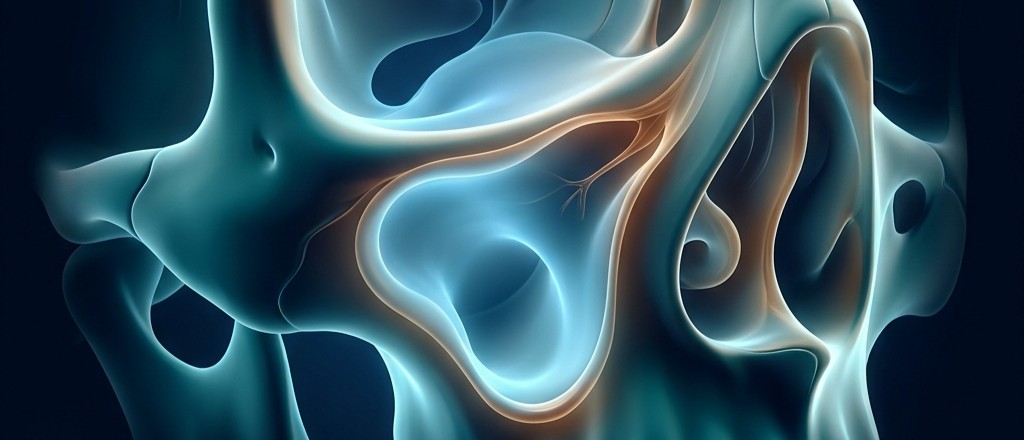
Anatomy of the maxillary sinus – and why it's important to us.
The maxillary sinus is the largest of the four paranasal cavities. It is located within the body of the maxilla and is lined by a thin respiratory mucosa known as the Schneiderian membrane, which in healthy individuals typically has a thickness of between 0.3 and 0.8 mm. Although it is extremely thin, this membrane possesses significant regenerative potential.
To plan the intervention, it is necessary to know the anatomy of the sinuses, including:
1. the height of the residual alveolar bone,
2. shape and volume of the sinus,
3. presence of bony septa (Underwood's septa),
4. position of the posterior superior alveolar artery,
5. the thickness of the lateral bony wall,
6. condition of the Schneider's membrane.
Why does bone loss occur after a tooth extraction?
Alveolar bone exists primarily to provide support for the teeth. After an extraction, its biological significance is almost lost, and the body begins the process of physiological resorption.
Data from the literature indicate that the greatest loss of bone volume occurs during the first six months after tooth extraction. During the first year, the width of the alveolar ridge can decrease by up to 50%, while the bone height gradually decreases in subsequent years, with no clear data on the rate of resorption.
What is a sinus lift?
A sinus lift is a surgical procedure that creates the conditions for further implant therapy in the lateral region of the upper jaw when a lack of bone and the sinus's anatomical position make it impossible. The procedure involves carefully elevating the Schneiderian membrane and creating a new space between the membrane and the bony floor of the sinus. This space is filled with a suitable augmentation material, which serves as the biological basis for the formation of new vital bone.
It is important to emphasize that a graft is not a permanent bone substitute. Its primary role is to provide a three-dimensional framework that allows for the migration of osteogenic cells, the formation of new blood vessels, and the gradual remodeling into vital bone tissue. Simply put, new bone is created where it is needed.
Thanks to these biological processes, the regenerated bone becomes capable of bearing the implant's functional load long-term after a few months.
When is a sinus lift indicated?
The decision to perform a sinus lift is made after a detailed clinical examination and analysis of the CBCT scan. The key parameter is the residual height of the alveolar bone.
Can every patient be a candidate for a sinus lift?
Although a sinus lift is a routine and very successful procedure, there are situations in which the intervention needs to be postponed or avoided.
Relative or absolute contraindications include active sinusitis, untreated odontogenic infections, uncontrolled diabetes, prior radiotherapy to the jaw region, the administration of intravenous bisphosphonates, severe coagulation disorders, as well as heavy smoking, which significantly increases the risk of unsuccessful regeneration. Of course, it is important to emphasize that the assessment is made by the prescribing physician, who, based on available information, consults with the physician treating the underlying condition. For this reason, it is conditionally defined in the literature as a relative contraindication.
Therefore, a detailed medical history as well as an essential physical examination are necessary before planning the surgery.
Lateral or crestal sinus lift – how is the appropriate technique chosen?
The selection is made by the surgeon, as expected, based on current recommendations and data from the literature.
International Team for Implantology (ITI) states that the choice between a lateral and a crestal sinus lift primarily depends on three factors that equally influence one another: the height of the alveolar bone, the possibility of achieving primary implant stability, and, of course, the anatomy of the maxillary sinus.
Crystalline sinus lift
A crestal or transalveolar sinus lift involves raising the sinus floor through the prepared opening for the future implant site, without forming a lateral bone window. The goal of the procedure is to increase the vertical height by a few millimeters to allow for the placement of an appropriately long implant without injuring the sinus membrane.
According to ITI recommendations, this technique is indicated for patients who have, most often, more than 5–6 mm of residual height.
The advantages of the crestal sinus lift include less surgical trauma, a shorter procedure time, less pronounced postoperative swelling, and the possibility of simultaneous implant placement in most cases. It is clear that the doctor will opt for this approach when anatomical conditions allow.
Lateral sinus lift
A lateral sinus lift involves accessing the maxillary sinus through an opening in its lateral wall.
The ITI consensus recommends this technique when the residual bone height is less than 5 mm, or when it is not possible to achieve adequate primary implant stability.
Implants can be placed during the same procedure or after a healing period of approximately six to nine months.
It is clear that the lateral sinus lift is more invasive because it involves greater manipulation of tissue, it is a longer procedure in the bone and consequently patient recovery is more difficult; however, its main advantage over the crestal approach is that it allows for significantly greater bone augmentation and represents the treatment of choice for severe atrophy in the posterior region of the upper jaw.
Which technique is better?
There is no single answer to this question, because, as with any surgical procedure, the therapy is based on indications. Therefore, for each individual case, the technique that overcomes anatomical deficiencies and results in a successful treatment is considered superior for that particular case.
Possible complications
The most common intraoperative complication of both techniques is perforation of the Schneiderian membrane, which is more frequent with the lateral approach due to the extent of the intervention.
The most common questions are whether this can be predicted and how it is resolved. For all surgical procedures, it is clear that everything is done with the goal of avoiding complications; however, if they do occur, it is important to know that the surgeon is familiar with the methods that lead to a solution. In this case, the solution is achieved by covering the perforation, most often with a collagen membrane, and the procedure continues.
A postoperative complication that is often a subject of discussion is sinus inflammation, or sinusitis. It is a rare complication and is most often the result of pre-existing sinonasal pathology, infection of the graft material used during surgery, or communication between the oral cavity and the sinus. Treatment in most cases involves the use of antibiotics, nasal decongestants, and saline solution. If this conservative approach does not lead to improvement, standard surgical treatment of the maxillary sinus is performed, which occurs in a smaller percentage of cases.
However, the standard complications that accompany any surgical procedure, in addition to those mentioned above, are certainly infection, swelling, pain, and bleeding, which are treated with well-established methods.
As already stated, proper diagnosis, careful planning, and adherence to surgical principles significantly reduce the risk of complications.
What does the postoperative course look like?
In the first days, there is moderate swelling, a feeling of pressure in the cheek area, and minimal nosebleeding.
Patients are advised to avoid blowing their nose, to sneeze with their mouths open, to avoid strenuous physical exertion, to avoid flying, and to regularly take their prescribed antibiotic and anti-inflammatory medications. It is very important to maintain oral hygiene, without skipping it for fear of injury.
Adherence to postoperative instructions directly impacts the success of the treatment. If these instructions are not followed, everything achieved through the doctor's careful work can be compromised, prolonging the treatment and leading to complications as well as additional surgical procedures.
Conclusion
Analysis of literature based on systematic reviews as well as longitudinal clinical studies has confirmed that implants placed after a successfully performed sinus lift achieve a survival rate of over 95% after ten years, which classifies this procedure as highly predictable with a high success rate.
It is important to emphasize that this article is informational and is intended for patients undergoing this therapeutic procedure, with an emphasis that therapy is individual for each patient.
The plan is formulated based on the clinical examination and medical history. The treatment protocol is determined by the oral surgeon based on clinical experience and expertise in the field. Therefore, if you have any questions, the only correct answer can be obtained during an examination from the doctor performing it.
Case presentation
The following is a clinical case that, through a series of X-ray images, follows the process from start to finish. The intervention includes Sinus lift with simultaneous implant placement, exclusively with augmentation patient's own (autologous) bone.
This approach is used when significant bone augmentation is not required (most often for a single implant), provided that the shape of the maxillary sinus is favorable and all walls are accessible.
The main advantage of using one's own bone is a drastically lower risk of infection and tissue inflammation compared to artificial materials. The procedure is more comfortable and cost-effective because it does not require the placement of a collagen membrane. Although the graft is naturally resorbed over time, in these indications, the regenerated bone is almost always sufficient for long-term success.

Insufficient bone in the region of the upper left first molar, several months after tooth extraction

Since the minimum length of an implant for placement in the upper jaw is 8mm, about 6mm is missing.

Recording after the intervention Sinus floor elevation with simultaneous by installing implant with a replacement of the patient's own bone.

After a healing period of at least six months, and often longer, a significant increase in the bone level of the subantral space is seen.

Final prosthetic restoration on an implant.